Description
FEATURES
- Designed for Deep Imaging
- Capture Fast, Dynamic Cellular Processes
- Spend Less Time Tuning the Laser
- Optional Features for Advanced Applications
- Intuitive Software Optimized for Multiphoton Observation
- Choose From 3 Frames and 3 Laser Configurations
- Modular Units Designed for Your Applications
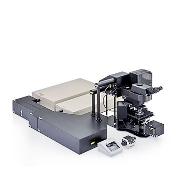
FLUOVIEW FVMPE-RS
| Laser Unit | IR Pulsed Laser with Negative Chirp for Multiphoton Excitation | <Spectra-Physics products> MAITAI HPDS-OL: 690 nm — 1040 nm MAITAI eHPDS-OL: 690 nm — 1040 nm INSIGHT X3-OL: 680 nm — 1300 nm INSIGHT X3 DUAL/DUALC-OL: 680 nm — 1300 nm + 1045 nm<Coherent products> Chameleon Vision I Olympus : 690 nm – 1040 nm Chameleon Vision II Olympus : 680 nm – 1080 nm Chameleon Vision S Olympus : 690 nm – 1050 nm<Main IR pulsed laser> One Laser System & Twin Lasers System: MAITAI HPDS-OL, MAITAI eHPDS-OL, INSIGHT X3-OL, Chameleon Vision I Olympus, Chameleon Vision II Olympus, Chameleon Vision S Olympus Dual Lines System:INSIGHT X3 DUAL-OL, INSIGHT X3 DUALC-OL <Additional IR line/ Laser> |
|||
|---|---|---|---|---|---|
| Laser Combiner | Introduction optic with AOM attenuation (0 % – 100 %, 0.1 % increment)Including fully automated beam expander, XY shifter and two axes angle alignment. (4 Axes Quadralign Auto Alignment optic) Direct coupling to laser port of scanning unit. <Dual Lines, Twin Lasers System> Motorized light path switcher, with DM900, DM1000R, DM1100 to combine two IR wavelength for imaging. |
||||
| Single laser for visible light without AOTF | [object Object] | ||||
| Scanning and Detection | Main Scanner | Standard Laser Ports |
|
||
| Detector | Standard |
|
|||
| Cooled GaAsP-PMT 2 CH |
|
||||
| Optional 4 CH |
|
||||
| Photo Detection Method | Analog Integration |
|
|||
| Galvanometer Scanner (Normal Imaging) | Galvanometer Mirror Scanner (X, Y) |
|
|||
| Scanning Modes | 2D | XY, XZ, XT, Xλ | |||
| 3D | XYZ, XYT, XYλ, XZT, XTλ, XZλ | ||||
| 4D | XYZT, XZTλ, XYTλ | ||||
| 5D | XYZTλ | ||||
| Other | ROI scanning: rectangle clip, ellipse, polygon, free area, line, free line and point | ||||
| Scanning Speed |
|
||||
| Scanning Zoom |
|
||||
| Resonant Scanner (High-Speed Imaging) | Scanning Modes | 2D | XY, XZ, XT | ||
| 3D | XYZ, XYT, XZT | ||||
| 4D | XYZT | ||||
| 5D | – | ||||
| other | ROI scanning: rectangle clip, line | ||||
| Scanning Speed | 30 fps at 512 x 512, 438 fps at 512 x 32. | ||||
| Scanning Zoom | 1.0 X – 8.0 X with 0.01 X increment | ||||
| Field Number (NA) |
|
||||
| Z-Drive |
|
||||
| Transmitted Light Detector Unit | Module with integrated external transmitted light photomultiplier detector and 100 W Halogen lamp, motorized switching, fi ber adaptation to microscope frame | ||||
| Microscope | Frame |
|
|||
| System Control | Controller |
|
|||
| Power Supply Unit | – | ||||
| Optional Unit | SIM Scanner |
|
|||
| Software | Basic Features | Dark room matching GUI design. User-arrangeable layout. Acquisition parameter reload features. Hard disk recording capability, adjust laser power and HV with Z-stack acquisition. Z-stack with alpha blending, maximum-intensity projection, iso-surface rendering. |
|||
| IR Laser Controlling |
|
||||
| Optional Motorised-Stage Software | XY motorised-stage control, map image acquisition for easy target locating. Tiling acquisition and software image stitching. Define multiple areas for time-lapse imaging. |
||||
| Optional Mapping and Multiplepoint Simulation Software | Multiple-point stimulation and data acquisition software. Mapping multiple-point stimulation to generate reaction map. Filtering feature to select points. | ||||
| Optional Sequencer Manager | Advanced programmable software to define multiple imaging/ stimulation tasks and execute by hardware sequencer. Minimum gap 100 ms delay between tasks. |
||||
| Optional Auto Compensation Software | Auto spherical aberration compensation software. Control of objective lens with auto spherical aberration compensation function. Auto adjustment of motorized correction collar to find the best position at certain observation depth. Auto adjustment of correction collar along with Z movement. |
||||
| Dimensions, Weight and Power Consumption | Microscope with Scan Unit | Dimensions (mm) |
|
||
| Operating Environment (Indoor Use) | Ambient Temperature | Room temperature: 20 – 25 ℃ | |||
| Maximum Relative Humidity | 75 % or less at 25 °C requires continuous (24-hour) power supply |
TruResolution Objectives
| FV30-AC10SV | FV30-AC25W | |
|---|---|---|
| Magnifications | 10 | 25 |
| NA | 0.6 | 1.05 |
| W.D. (mm) | 8 | 2 |
| Cover Glass Thickness (mm) | 0 – 0.23 | 0 – 0.23 |
| Immersion Liquid | SCALEVIEW-A2 (Water, Silicone oil, and Normal oil available) | Water |
| Special features | Auto Compensation, Optimized for multiphoton imaging | Auto Compensation, Optimized for multiphoton imaging |
| Dimensions | 56mm (W) x 106.5mm (D) x 95mm(H)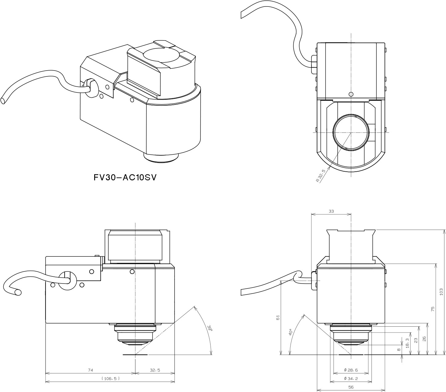 |
56mm (W) x 106.5mm (D) x 101mm(H)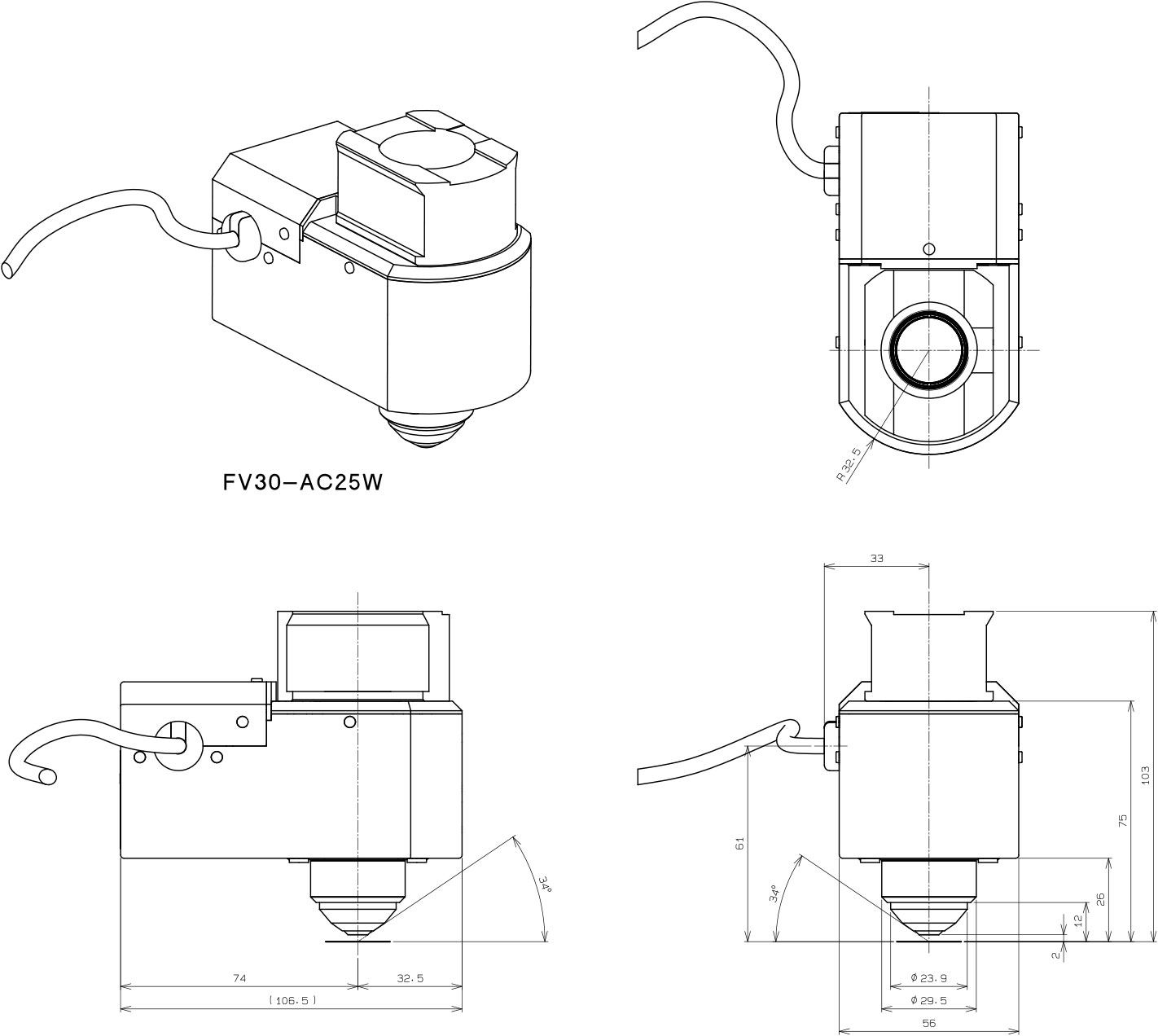 |
| Weight | approx. 1kg | approx. 1kg |